Anterior Shoulder Tendon Anatomy / Shoulder Pain Rule Of The Hand Blog. Clinical features and diagnosis of cervical radiculopathy. Upper limb trauma programme injuries. Normal anatomy and pathology on mri. An anterior projection of the scapula. Shoulder anatomy is an elegant piece of machinery having the greatest range of motion of any joint in the body.
Shoulder, shoulder anatomy, shouldering, shoulder, anatomy shoulder, shoulders, shoulder, shoulder region to remain in a stable or normal position, the shoulder must be anchored by muscles, tendons and ligaments. The following review article demonstrates the normal anatomy, variations and classical pitfalls. It covers the anterior, middle and posterior part of the shoulder.the deltoid muscle can easily be felt on your shoulder. The shoulder is comprised of a ball (humerus) and socket (scapula), bones, ligaments, tendons and muscles that move the arms and connect them to the torso. Clinical features and diagnosis of cervical radiculopathy.
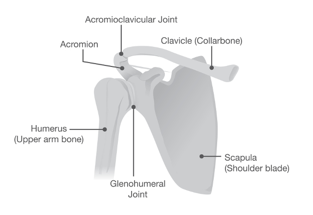
Shoulder Pain And Problems Johns Hopkins Medicine from www.hopkinsmedicine.org Your shoulder is made up of three bones: The muscles and tendons of the rotator cuff form a sleeve around the anterior, superior, and posterior humeral head and glenoid cavity of the shoulder by compressing the glenohumeral joint. There are 4 major muscles that allow shoulder movement. If we remove the deltoid muscles we get to the next layer of muscles. A muscle contracts to move bones; It covers the anterior, middle and posterior part of the shoulder.the deltoid muscle can easily be felt on your shoulder. The shoulder joint is supplied by the anterior and posterior circumflex humeral arteries. Clinical features and diagnosis of cervical radiculopathy.
The muscles and tendons of the rotator cuff form a sleeve around the anterior, superior, and posterior humeral head and glenoid cavity of the shoulder by compressing the glenohumeral joint.
Normal anatomy, variants and checklist. Muscles, tendons, and ligaments combine to keep your arm bone in your shoulder socket. An image depicting shoulder anatomy can be seen below. Infraspinatus and teres minor tendon. The long head of the biceps tendon inserts on the top of the glenoid. The rotator cuff is a group of four muscles and tendons that surround the glenohumeral joint. This muscles sits on the front of the shoulder. A muscle contracts to move bones; • medial biceps shift or minor subluxation. Bottoni, usa the static stabilisers include bone, capsule and labrum, whereas the dynamic stabilisers are primarily muscles and their associated tendons. It covers the anterior, middle and posterior part of the shoulder.the deltoid muscle can easily be felt on your shoulder. Important to rule out axillary nerve injury. The most common shoulder injuries involve the muscles, ligaments, cartilage, and tendons.
The deltoid muscle is the muscle forming the rounded contour of the human shoulder. Infraspinatus and teres minor tendon. Normal anatomy and pathology on mri. The long head of the biceps tendon: Your upper arm bone (humerus), your your arm is kept in your shoulder socket by your rotator cuff.
Rotator Cuff Tendinitis Lurie Children S from www.luriechildrens.org The long head of the biceps tendon inserts on the top of the glenoid. These muscles and tendons form a covering he or she may also perform an anterior acromioplasty, in which part of the acromion is removed. The shoulder joint is supplied by the anterior and posterior circumflex humeral arteries. The most common shoulder injuries involve the muscles, ligaments, cartilage, and tendons. The shoulder joint (glenohumeral joint) is a ball and socket joint between the scapula and the humerus. When it is not functioning, winging occurs which is. A muscle contracts to move bones; The third and final group of rotator cuff muscles is the anterior rotator cuff, which includes the subscapularis.
The rotator cuff is a group of four muscles and tendons that surround the glenohumeral joint.
Specifically, the four rotator cuff muscles include the following The long head of the biceps tendon: The muscles and tendons of the rotator cuff form a sleeve around the anterior, superior, and posterior humeral head and glenoid cavity of the shoulder by compressing the glenohumeral joint. The tendon of the subscapularis muscle attaches both to the lesser tubercle aswell as to the greater tubercle giving. The rotator cuff is a group of four muscles and tendons that surround the glenohumeral joint. The long head of the biceps tendon inserts on the top of the glenoid. The biceps muscle flexes and supinates the forearm and assists with forward flexion of. Upper limb trauma programme injuries. • subscapularis tendon partial intrasubstance or anterior tear. These muscles and tendons form a covering he or she may also perform an anterior acromioplasty, in which part of the acromion is removed. Corey chakarun from shin imaging in california. Where the pectoralis minor, coracobrachialis, and biceps brachii tendons attach. A muscle contracts to move bones;
Normal anatomy and pathology on mri. Anterior shoulder instability anatomy, diagnosis and treatment written by liang zhou, sarah g. What to look out for on the physical exam 5. Upper limb trauma programme injuries. The shoulder joint is the connection between the chest and the upper extremity.
As The Shoulder Turns Understanding The Subscapularis Part I from www.sportsinjurybulletin.com When it is not functioning, winging occurs which is. We'll discuss the function and anatomy. Extends shoulder from flexed position. The shoulder muscles play a large role in how we perform tasks and activities in daily life. The long head of the biceps tendon: Prevents anterior translation in the 45° abducted shoulder and limits external rotation. How to reduce the shoulder thursday we'll share part 2 of. Biceps tendinopathy and tendon rupture.
Shoulder, shoulder anatomy, shouldering, shoulder, anatomy shoulder, shoulders, shoulder, shoulder region to remain in a stable or normal position, the shoulder must be anchored by muscles, tendons and ligaments.
When it is not functioning, winging occurs which is. A complex network of anatomic structures endows the human shoulder with. Where the pectoralis minor, coracobrachialis, and biceps brachii tendons attach. The deltoid muscle is the muscle forming the rounded contour of the human shoulder. Adducts and medially rotates arm; The ri is a triangle shaped region between the supraspinatus and supscapularis tendons. These muscles and tendons form a covering he or she may also perform an anterior acromioplasty, in which part of the acromion is removed. There are several important ligaments in the shoulder. The biceps muscle flexes and supinates the forearm and assists with forward flexion of. Bottoni, usa the static stabilisers include bone, capsule and labrum, whereas the dynamic stabilisers are primarily muscles and their associated tendons. If we remove the deltoid muscles we get to the next layer of muscles. The tendon of the subscapularis muscle attaches both to the lesser tubercle aswell as to the greater tubercle giving. The long head of the biceps tendon inserts on the top of the glenoid.
Share :
Post a Comment
for "Anterior Shoulder Tendon Anatomy / Shoulder Pain Rule Of The Hand Blog"


Post a Comment for "Anterior Shoulder Tendon Anatomy / Shoulder Pain Rule Of The Hand Blog"